Richard L. Drake & A. Wayne Vogl & Adam W. M. Mitchell & Richard Tibbitts & Paul Richardson
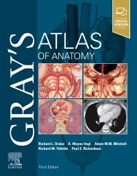
Clinically focused, consistently and clearly illustrated, and logically organized, Gray's Atlas of Anatomy, the companion resource to the popular Gray's Anatomy for Students, presents a vivid, visual depiction of anatomical structures. Stunning illus
...view more
Clinically focused, consistently and clearly illustrated, and logically organized, Gray's Atlas of Anatomy, the companion resource to the popular Gray's Anatomy for Students, presents a vivid, visual depiction of anatomical structures. Stunning illustrations demonstrate the correlation of structures with clinical images and surface anatomy - essential for proper identification in the dissection lab and successful preparation for course exams.
Clinically focused, consistently and clearly illustrated, and logically organized, Gray's Atlas of Anatomy, the companion resource to the popular Gray's Anatomy for Students, presents a vivid, visual depiction of anatomical structures. Stunning illustrations demonstrate the correlation of structures with clinical images and surface anatomy - essential for proper identification in the dissection lab and successful preparation for course exams.
Key Features
Build on your existing anatomy knowledge with structures presented from a superficial to deep orientation, representing a logical progression through the body.
Identify the various anatomical structures of the body and better understand their relationships to each other with the visual guidance of nearly 1,000 exquisitely illustrated anatomical figures.
Visualize the clinical correlation between anatomical structures and surface landmarks with surface anatomy photographs overlaid with anatomical drawings.
Recognize anatomical structures as they present in practice through more than 270 clinical images - including laparoscopic, radiologic, surgical, ophthalmoscopic, otoscopic, and other clinical views - placed adjacent to anatomic artwork for side-by-side comparison.
Gain a more complete understanding of the inguinal region in women through a brand-new, large-format illustration, as well as new imaging figures that reflect anatomy as viewed in the modern clinical setting.
Enhanced eBook version included with purchase. Your enhanced eBook allows you to access all of the text, figures, and references from the book on a variety of devices – as well as dissection videos and self-assessment questions and answers.
Author Information
By Richard L. Drake, PhD, Director of Anatomy, Professor of Surgery, Cleveland Clinic Lerner College of Medicine, Case Western Reserve University, Cleveland, Ohio; A. Wayne Vogl, PhD, Professor of Anatomy & Cell Biology, Department of Cellular and Physiological Sciences, Faculty of Medicine, University of British Columbia, Vancouver, British Columbia, Canada;; Adam W. M. Mitchell, MB BS, FRCS, FRCR, Lecturer, Interventional Fellow, Department of Interventional Radiology, Hammersmith Hospital, London, UK; Richard Tibbitts and Paul Richardson, Cambridge, UK
https://www.us.elsevierhealth.com/grays-atlas-of-anatomy-9780323636391.html3564Gray's Atlas of Anatomyhttps://www.us.elsevierhealth.com/media/catalog/product/9/7/9780323636391.jpg84.9984.99USDInStock/Medicine/Anatomy/Medical Students/Anatomy/Books12135255039<P>Clinically focused, consistently and clearly illustrated, and logically organized, <I>Gray's Atlas of Anatomy</I>, the companion resource to the popular <I>Gray's Anatomy for Students</I>, presents a vivid, visual depiction of anatomical structures. Stunning illustrations demonstrate the <B>correlation of structures with clinical images and surface anatomy</B> - essential for proper identification in the dissection lab and successful preparation for course exams.</P> <P>Clinically focused, consistently and clearly illustrated, and logically organized, <I>Gray's Atlas of Anatomy</I>, the companion resource to the popular <I>Gray's Anatomy for Students</I>, presents a vivid, visual depiction of anatomical structures. Stunning illustrations demonstrate the <B>correlation of structures with clinical images and surface anatomy</B> - essential for proper identification in the dissection lab and successful preparation for course exams.</P>00add-to-cart97803236363912020ProfessionalBy Richard L. Drake, PhD, A. Wayne Vogl, PhD, Adam W. M. Mitchell, MB BS, FRCS, FRCR, Richard Tibbitts and Paul Richardson20213Book216w x 276h1000 illustrations (1000 in full color)Churchill Livingstone648Feb 17, 2020IN STOCKBy <STRONG>Richard L. Drake</STRONG>, PhD, Director of Anatomy, Professor of Surgery, Cleveland Clinic Lerner College of Medicine, Case Western Reserve University, Cleveland, Ohio; <STRONG>A. Wayne Vogl</STRONG>, PhD, Professor of Anatomy & Cell Biology, Department of Cellular and Physiological Sciences, Faculty of Medicine, University of British Columbia, Vancouver, British Columbia, Canada;; <STRONG>Adam W. M. Mitchell</STRONG>, MB BS, FRCS, FRCR, Lecturer, Interventional Fellow, Department of Interventional Radiology, Hammersmith Hospital, London, UK; <STRONG>Richard Tibbitts</STRONG> and <STRONG>Paul Richardson</STRONG>, Cambridge, UKBooks, eBooksBooksGray's AnatomyNoNoNoNoPlease SelectPlease SelectPlease Select